News Article
Radiation Safety Expert Strikes Gold in International Competition
News Story from 18th May 2016
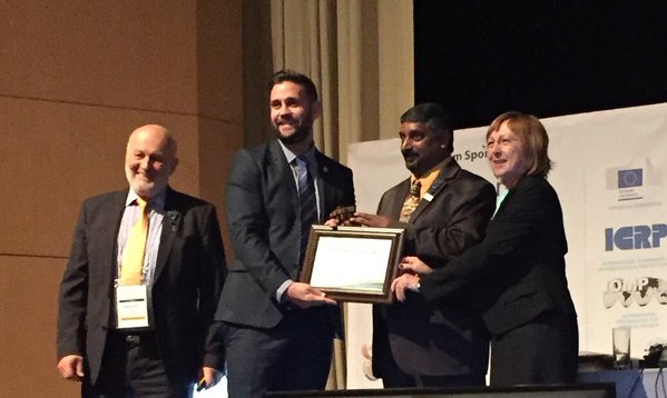
A medical physicist’s work on how to get better quality X-ray images of babies’ ankles without increasing the radiation dose has received international recognition from his peers. Adam Jones, 30, who developed the technique while working for the Guy’s & St Thomas’ NHS Foundation Trust in London, won the International Radiation Protection Association’s (IRPA) Young Scientist and Professionals Award Competition. His prize was a certificate of achievement and a solid gold Kruger rand, a fitting choice for the South African setting. Twenty young scientists from around the world, representing their national organisations promoting radiation safety, competed at IRPA’s four yearly Congress held recently in Cape Town, South Africa. Adam presented his findings to an international panel of judges and other delegates during the week long conference. He had been selected to represent the UK by the Society for Radiological Protection (SRP), after winning a national competition in 2014. The fourteen-strong UK delegation was led by SRP President Professor Pete Cole of Liverpool University. When results were revealed during the closing ceremony Professor Cole said “Adam and the UK profession should feel extremely proud of this win. The award is only made every four years. This is a wonderful achievement against outstanding competitors representing the crème of the younger radiation protection professionals. SRP and our partner societies salute Adam’s expertise and application”.
Just why is the work so important? “Sometimes X-ray images of babies’ ankles were not good enough quality to help make decisions about their care,” said Adam. “We couldn’t just increase the radiation dose to get a better image because UK regulations say patients’ exposure to radiation should be as low as possible whilst still getting a useable picture, so first we had to check whether there was any other way we could improve the image quality.” Adam tried setting up the equipment in different ways and took images of a ‘phantom’ baby’s foot, a model that trainee radiographers practice on. He found that one particular set up had significantly improved the image quality. Adam believes this technique could also be used to improve X-ray image quality of other parts of the body such as hands, feet and elbows. “Because this new process uses existing equipment it means we don’t have to invest anything extra and it’s quick to put into practice” he said. Since 2014, Adam’s work has been published in the British Journal of Radiology and the technique is being adopted throughout the UK and Europe. It has become a case study example on optimisation within European Training and Education for Medical Physics Experts in Radiology (EUtempe).
Adam relocated to the Medical Physics Department, Western Sydney Local Health District, Sydney, New South Wales, in October 2014, from where he hopes this important improvement will be rolled out across Australia and continue to be adopted on a global scale.
- It's First Place for UK's Adam Jones in… 13th May 2016
Women in Radiation Protection at the Highest Level
Posted 13th May 2016
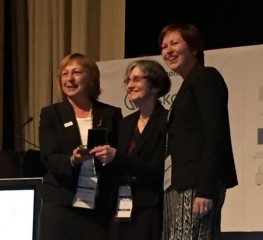
At this morning's IRPA14 Plenary Session in Cape Town the Gold Medal for Radiation Protection was…